How Is It Treated?
At birth, the baby with TGA is usually stable. However careful examination often reveals that there is some degree of cyanosis (blueness). If the ductus arteriosis begins to close or if the Foramen Ovale becomes restrictive, then the infant can show signs of worsening cyanosis and respiratory distress. Drugs (such as prostaglandin E1) may make the infant less blue by causing the ductus arteriosus, a small blood vessel that connects the aorta with the pulmonary artery, to reopen.
If oxygen levels are still too low after prostaglandin therapy, or if the infant is very ill, temporary relief may be achieved by performing a procedure called a Balloon Septostomy (see animation at left) in the catheterization lab.
The Balloon Septostomy involves the insertion of a tube (catheter) with an uninflated balloon at its end from a leg vein into the heart and through the foramen ovale into the left atrium. The balloon is then inflated and withdrawn, tearing the atrial septum as it is pulled back into the right atrium. This large opening allows for better mixing of the unoxygenated (blue) and oxygenated (red) blood.
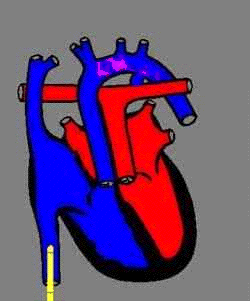
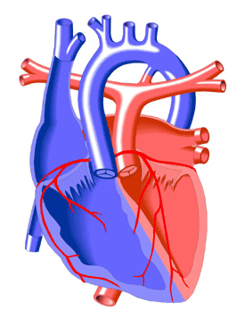
A Balloon Septostomy in the catheterization lab is only a temporary measure to stabilize the neonate, however. When the infant is about one week old, an Arterial Switch Operation is performed which corrects the defect by connecting the aorta to the left ventricle and the pulmonary artery to the right ventricle, as in a normal heart (see animation at right).
A component of this surgical repair involves reconnecting the small coronary arteries to the aorta so that the heart muscle receives red (oxygenated) blood. The foramen ovale is also closed at this time and the patent ductus arteriosus (PDA) is closed off. |